Before and After 2010: Treatment of Cleft Lip and Palate – a “Pro-bono” Smile Makeover

A female patient in her early 20s presented with congenitally missing upper lateral incisors, and one canine, a total of 3 teeth seeking a fixed replacement. The patient has a cleft lip and palate bilaterally, which has been treated several times over the years with surgery to repair it.
Implants are not possible as there is no bone in the surgical repair sites, and in fact, she still has an oro-nasal communication.
The patient had limited resources to pay for treatment and hoped that the federal Cleft Lip and Palate Scheme would cover the costs, which turned out that only extractions and dentures are eligible for the claim. Thus, the treating clinician – Dr Michael Mandikos and I had decided to treat the case at no cost – pro-bono – with 3M ESPE’s support.
We aimed to set up for a somewhat ‘experimental’ application of Zirconia as a frame material for bonded ‘Maryland Style’ bridges, with refractory veneers for the rest to maintain the same property in the layering ceramics.


Figs 1-3 show the retracted views of pre-op condition. Note the large gap between the central (21) and canine (23) on the left.
Fig 5 shows the Computerised Tomography (CT) Scan image of the pre-op condition to assess detailed hard-tissue information.
Because of the limited option in replacing teeth while preserving the virgin dentitions as much as possible, the patient agreed to proceed with orthodontic treatment for 18 months to minimise the clinical and technical difficulties (Fig 4 & 6).


Fig 7 shows the completed model work for the refractory veneers and ceramic build for the Maryland bridges to replace the laterals. Fig 8 shows the image captured during the degassing of the refractory dies.
Fig 9 shows the first dentin porcelain/ceramic build-up, trying to establish the internal structure in the right position. Fig 10 shows the completed restorations on the model.

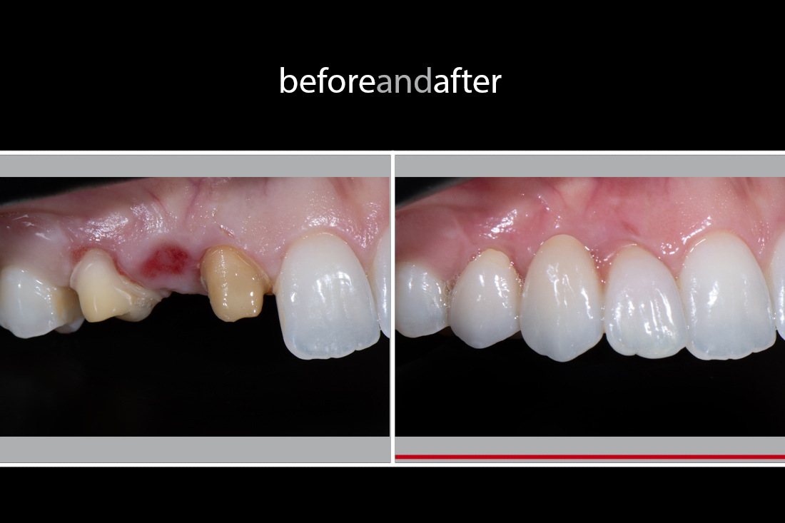
Figs 11 and 12 show the post-op images captured in June 2010. Note the effect of the orthodontic treatment closing the gap in Q2.

Figs 13 and 14 show the post-op images captured in August 2020, a 10-year follow-up. Everything is still intact though some ortho relapse, most notably in the difference in the incisal edge at the central incisors.
As mentioned at the beginning of this post, this was an experimental case using zirconia for the Maryland bridges (typically lithium disilicate is the standard material for the bonding strength); however, the result serves as clinical evidence and it works just fine as shown above. The cement was RelyX Veneer cement combined with SingleBond 2. Dr Mandikos has been using this technique for a decade and claims no technical and bonding strength issues.
Technically speaking, if I were to receive the case now in 2021, I would lean towards using lithium disilicate for the following reasons:
- to reduce the difficulty of the case
- to achieve a more predictable outcome and better control during the ceramic build-up – the refractory veneers were over 2mm in thickness incisally, which was challenging to determine the ideal incisal edge positions caused by its significant ceramic shrinkage
- better strength (of the veneers) for peace of mind.
Having said, I feel that we, of course, successfully achieved a great outcome in a challenging situation with longevity (at least lasing ten years so far). This case serves as good evidence to apply where the bridges’ connector dimensions are limiting, and intricate colour matching is required (as I find Zirconia layering ceramic has better control than lithium disilicate’s).

Fig 15 shows the post-op full-face photo received from the patient back in 2010, and she said:
“ I have not stopped smiling for what feels like decades now. Yugo and Michael have changed my life, and I will be forever grateful with everything you both have done for me”.
Acknowledgement:
I would like to thank Dr Michael Mandikos from Brisbane Prosthodontics in Brisbane QLD, Australia, for his clinical excellence, and the photographs. https://www.brisbaneprosthodontics.com.au/
I would also like to thank 3M ESPE for being a sponsor / a part of the experimental research, and providing the necessary clinical and technical materials.
This case was issued in June 2010 with no remakes.
Reference:
Sahoo NK, Desai AP, Roy ID, Kulkarni V. Oro-Nasal Communication. J Craniofac Surg. 2016 Sep;27(6):e529-33